
Mouse Secondary Antibodies

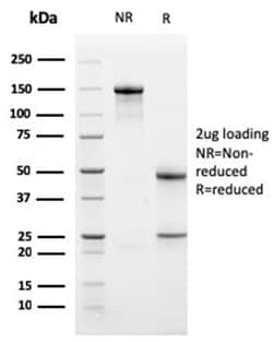
Biotium Bcl-X (Apoptosis Marker)(BCL2L1/1762), CF568 conjugate, 0.1mg/mL
Bcl-x belongs to the Bcl-2 family of proteins and plays a crucial role in apoptosis. Bcl-X has two isoforms: Bcl-XL (long), a 26 kDa protein which suppresses cell death, and Bcl-XS (short), a 178 amino acid protein lacking a 63 amino acid domain which functions as a dominant inhibitor of Bcl-2 and promotes apoptosis. This MAb reacts with both Bcl-XS and Bcl-XL proteins, and shows no cross-reaction with Bcl-2 or Bax proteins.Primary antibodies are available purified, or with a selection of fluorescent CF Dyes and other labels. CF Dyes offer exceptional brightness and photostability. Note: Conjugates of blue fluorescent dyes like CF405S and CF405M are not recommended for detecting low abundance targets, because blue dyes have lower fluorescence and can give higher non-specific background than other dye colors.

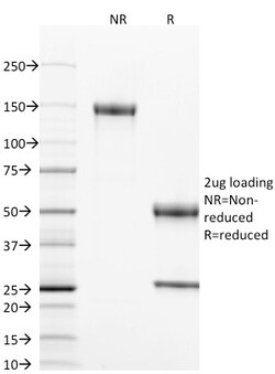
Biotium CD40 / TNFRSF5 / CD40L-Receptor(T8P2G4*A6), CF568 conjugate, 0.1mg/mL
This antibody is specific for the soluble form of CD40. It is a receptor on antigen-presenting cells of the immune system and is essential for mediating a broad variety of immune and inflammatory responses including T cell-dependent immunoglobulin class switching, memory B cell development, and germinal center formation. AT-hook transcription factor AKNA is reported to coordinately regulate the expression of this receptor and its ligand, which may be important for homotypic cell interactions. Adaptor protein TNFR2 interacts with this receptor and serves as a mediator of the signal transduction. The interaction of this receptor and its ligand is found to be necessary for amyloid-beta-induced microglial activation, and thus is thought to be an early event in Alzheimer disease pathogenesis. CD40 is expressed on B-lymphocytes, follicular dendritic cells, bone marrow-derived dendritic cells, thymic epithelium, and interdigitating cells in the T-cell zones of secondary lymphoid org
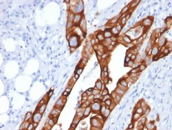
Biotium Cytokeratin 20 (KRT20) (Colorectal Epithelial Marker)(KRT20/1991), 1mg/mL
This MAb recognizes an intermediate filament protein of 46 kDa, identified as cytokeratin 20 (KRT20). KRT is abundantly expressed in goblet cells and enterocytes of the gastrointestinal tract. It is a useful marker of pancreatic and colorectal cancer. KRT20 is expressed under normal, hyperplastic and neoplastic conditions. It has been detected in adenocarcinomas of the colon, stomachand biliary tract. Breast carcinomas are generally non-reactive. Primary antibodies are available purified, or with a selection of fluorescent CF Dyes and other labels. CF Dyes offer exceptional brightness and photostability. Note: Conjugates of blue fluorescent dyes like CF405S and CF405M are not recommended for detecting low abundance targets, because blue dyes have lower fluorescence and can give higher non-specific background than other dye colors.

Biotium TIMP2 (Tissue Inhibitor of Metalloproteinase 2) (3A4), CF568 conjugate, 0.1mg/mL
This antibody recognizes a protein of 21 kDa, identified as tissue inhibitor of metalloproteinases-2 (TIMP-2). It is closely related to TIMP-1 and shows the highest binding affinity to both the latent (pro) and active forms of 72 kDa Type IV collagenase (also known as MMP-2 or gelatinase A). It also has affinity for the active form of 92 kDa Type IV collagenase (also known as MMP-9 or gelatinase B). TIMP-2 inhibits the proteolytic invasiveness of tumor cells and normal placental trophoblast cells.Primary antibodies are available purified, or with a selection of fluorescent CF Dyes and other labels. CF Dyes offer exceptional brightness and photostability. Note: Conjugates of blue fluorescent dyes like CF405S and CF405M are not recommended for detecting low abundance targets, because blue dyes have lower fluorescence and can give higher non-specific background than other dye colors.
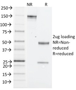
Biotium NK1.1 / CD161c / Klrb1c, Mouse(PK136), CF568 conjugate, 0.1mg/mL
NK1. 1 is a type II integral membrane glycoprotein with a C-type lectin domain and is encoded by theKlrb1c/NKR-P1Cgene. It is predominantly expressed as a disulfide-linked homodimer on NK cells however; it is also expressed on NK-T cells, a rare population of T lymphocytes. NK1. 1 is expressed in some mouse strains, including C57BL/6, FVB/N, and NZB, but not AKR, BALB/c, CBA/J, C3H, DBA/1, DBA/2, NOD, SJL, and 129. Expression of NKR-P1C antigen has been correlated with lysis of tumor cells in vitro and rejection of bone marrow allograftsin vivo. NK-1. 1 has also been shown to play a role in NK cell activation, IFN- production, and cytotoxic granule release. NK-1. 1 is commonly used as a mouse NK cell marker. Primary antibodies are available purified, or with a selection of fluorescent CF Dyes and other labels. CF Dyes offer exceptional brightness and photostability. Note: Conjugates of blue fluorescent dyes like CF405S and CF405M are not recommended for detecting low abu
Biotium Galectin-1 / Human Placental Lactogen (hPL) (GAL1/1831), 0.2mg/mL
Galectin-1 is a member of the beta-galactoside-binding family and is a dimeric protein of 14 kD participating in a variety of normal and pathological processes, including cancer progression. Galectin-1 can affect the proliferation of normal and malignant cells. Inhibition of cell growth is observed in a lactose-dependent manner as lower concentrations of the lectin stimulate cell proliferation. Galectin-1 may also be implicated in the induction of apoptosis of activated T cells through the binding of exogenous galectin-1 to CD45 molecules present on the surface of lymphocytes. Galectin-1, reported to be present either at the surface of cancer cells or accumulated around these cells could act as an immunological shield to protect against a T cell immune response and provide an advantage for survival.Primary antibodies are available purified, or with a selection of fluorescent CF Dyes and other labels. CF Dyes offer exceptional brightness and photostability. Note: Conjugates
Biotium S100A (Melanoma Marker) (S1/61), 0.2mg/mL
S100A and S100B proteins are two members of the S100 family of calcium binding proteins. S100A is composed of an alpha and a beta chain whereas S100B is composed of two beta chains. This antibody is specific against an epitope located on the alpha-chain (i.e. in S-100A and S-100B) but not on the beta-chain of S-100 (i.e. in S-100B). This antibody can be used to localize S-100A in various tissue sections. S-100 protein has been found in normal melanocytes, Langerhans cells, histiocytes, chondrocytes, lipocytes, skeletal and cardiac muscle, epithelial and myoepithelial cells of the breast, salivary and sweat glands. Neoplasms derived from these cells also express S-100 protein. Almost all malignant melanomas and cases of histiocytosis X are positive for S-100 protein.Primary antibodies are available purified, or with a selection of fluorescent CF Dyes and other labels. CF Dyes offer exceptional brightness and photostability. Note: Conjugates of blue fluorescent dyes like CF
Biotium Insulin / IRDN (beta-Cell & Insulinoma Marker) (rIRDN/805), CF568 conjugate, 0.1mg/mL
This antibody recognizes a polypeptide which is identified as insulin, a 51-amino acid polypeptide composed of A and B chains connected through the C-peptide. Proinsulin, which has very little biological activity, is cleaved by proteases within its cell of origin into the insulin molecule and the C-terminal basic residue. Insulin enhances membrane transport of glucose, amino acids, and certain ions. It also promotes glycogen storage, formation of triglycerides, and synthesis of proteins and nucleic acids. Deficiency of insulin results in diabetes mellitus. The main storage site for insulin is the pancreatic islets. Antibodies to insulin are important as beta-cell and insulinoma markers.Primary antibodies are available purified, or with a selection of fluorescent CF Dyes and other labels. CF Dyes offer exceptional brightness and photostability. Note: Conjugates of blue fluorescent dyes like CF405S and CF405M are not recommended for detecting low abundance targets, because
Biotium ZFYVE28 (Zinc Finger FYVE-type containing 28)(LST2/2426), CF647 conjugate, 0.1mg/mL
ZFYVE28 belongs to the lst-2 family. It contains 1 FYVE-type zinc finger. The FYVE-type zinc finger mediates the interaction with phosphatidylinositol 3-phosphate (PI3P) and localization to early endosome membranes when not mono-ubiquitinated at Lys-87. Mono-ubiquitination at Lys-87 prevents binding to phosphatidylinositol 3-phosphate (PI3P) and localization to early endosome membranes. ZFYVE28 is a negative regulator of epidermal growth factor receptor (EGFR) signaling. It acts by promoting EGFR degradation in endosomes when not mono-ubiquitinated. The FYVE domain has been identified in a number of unrelated signaling molecules. This protein functions to recruit SMAD2 to the transforming growth factor-beta receptor. The FYVE domain is required to maintain the normal localization of this protein but is not involved in mediating interaction with SMADs. Primary antibodies are available purified, or with a selection of fluorescent CF Dyes and other labels. CF Dyes offer except
Biotium S100A (Melanoma Marker) (S1/61), CF488A conjugate, 0.1mg/mL
S100A and S100B proteins are two members of the S100 family of calcium binding proteins. S100A is composed of an alpha and a beta chain whereas S100B is composed of two beta chains. This antibody is specific against an epitope located on the alpha-chain (i.e. in S-100A and S-100B) but not on the beta-chain of S-100 (i.e. in S-100B). This antibody can be used to localize S-100A in various tissue sections. S-100 protein has been found in normal melanocytes, Langerhans cells, histiocytes, chondrocytes, lipocytes, skeletal and cardiac muscle, epithelial and myoepithelial cells of the breast, salivary and sweat glands. Neoplasms derived from these cells also express S-100 protein. Almost all malignant melanomas and cases of histiocytosis X are positive for S-100 protein.Primary antibodies are available purified, or with a selection of fluorescent CF Dyes and other labels. CF Dyes offer exceptional brightness and photostability. Note: Conjugates of blue fluorescent dyes like CF
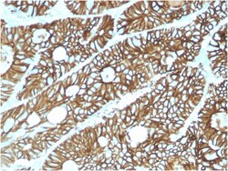
Biotium Cadherin 17 / LI Cadherin (Liver-Intestine Marker)(CDH17/2615), CF543 conjugate, 0.1mg/mL
Cadherin 17, also known as LI cadherin (for liver-intestine cadherin), is a member of the cadherin family of calcium-dependent adhesion molecules. Cadherins mediate cell-cell interactions via hemophilic binding, and are critical to the maintenance of tissue structure and morphogenesis. Cadherins each contain a large extracellular domain at the amino terminus, which is characterized by a series of five homologous repeats, the most distal of which is thought to be responsible for binding specificity. The relatively short carboxy terminal, intracellular domain interacts with a variety of cytoplasmic proteins, including beta-catenin, to regulate cadherin function. Cadherin 17 expression is restricted to liver and intestine tissues and is specifically localized to the basolateral domain of hepatocytes and enterocytes. Primary antibodies are available purified, or with a selection of fluorescent CF Dyes and other labels. CF Dyes offer exceptional brightness and photostability. N
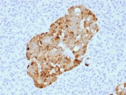
Biotium Chromogranin A / CHGA (Neuroendocrine Marker) (CHGA/1731R), CF488A conjugate, 0.1mg/mL
Chromogranin A is present in neuroendocrine cells throughout the body, including the neuroendocrine cells of the large and small intestine, adrenal medulla and pancreatic islets. It is an excellent marker for carcinoid tumors, pheochromocytomas, paragangliomas, and other neuroendocrine tumors. Co-expression of chromogranin A and neuron specific enolase (NSE) is common in neuroendocrine neoplasms. Reportedly, co-expression of certain keratins and chromogranin indicates neuroendocrine lineage. The presence of strong anti-chromogranin staining and absence of anti-keratin staining should raise the possibility of paraganglioma. The co-expression of chromogranin and NSE is typical of neuroendocrine neoplasms. Most pituitary adenomas and prolactinomas readily express chromogranin.Primary antibodies are available purified, or with a selection of fluorescent CF Dyes and other labels. CF Dyes offer exceptional brightness and photostability. Note: Conjugates of blue fluorescent dyes
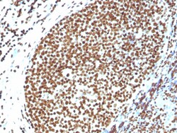
Biotium Histone H1 (Pan Nuclear Marker)(N/A), CF488A conjugate, 0.1mg/mL
Eukaryotic histones are basic and water-soluble nuclear proteins that form hetero-octameric nucleosome particles by wrapping 146 base pairs of DNA in a left-handed super-helical turn sequentially to form chromosomal fiber. Two molecules of each of the four core histones (H2A, H2B, H3, and H4) form the octamer; formed of two H2A-H2B dimers and two H3-H4 dimers, forming two nearly symmetrical halves by tertiary structure. Over 80% of nucleosomes contain the linker Histone H1, derived from an intronless gene that interacts with linker DNA between nucleosomes and mediates compaction into higher order chromatin. Histones are subject to posttranslational modification by enzymes primarily on their N-terminal tails, but also in their globular domains. Such modifications include methylation, citrullination, acetylation, phosphorylation, sumoylation, ubiquitination and ADP-ribosylation. Primary antibodies are available purified, or with a selection of fluorescent CF Dyes and other lab
Biotium Spectrin Alpha 1 (Erythrocyte Marker) (SPTA1/1810), CF488A conjugate, 0.1mg/mL
Spectrin is an actin crosslinking and molecular scaffold protein that links the plasma membrane to the actin cytoskeleton, and functions in the determination of cell shape, arrangement of transmembrane proteins, and organization of organelles. Spectrins function as membrane organizers and stabilizers, composed of non-homologous α andβ chains, which aggregate side-to-side in an anti-parallel fashion to form dimers, tetramers and higher polymers. Spectrin α I and spectrinβ I are present in erythrocytes, whereas spectrin α II (also designated fodrin α) and spectrinβ II (also designated fodrinβ) are present in other somatic cells. The spectrin tetramers in erythrocytes act as barriers to lateral diffusion, but spectrin dimers seem to lack this function.Primary antibodies are available purified, or with a selection of fluorescent CF Dyes and other labels. CF Dyes offer exceptional brightness and photostability. Note: Conjugates of blue fluorescent dyes like CF405S
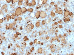
Biotium Thyroglobulin (Thyroidal Cell Marker) (rTGB24), CF647 conjugate, 0.1mg/mL
Thyroglobulin is a 660 kDa dimeric pre-protein with multiple glycosylation sites. It is produced by and processed within the thyroid gland to produce the hormone thyroxine and triiodothyronine. Prior to forming dimers, thyroglobulin monomers undergo conformational maturation in the endoplasmic reticulation. The vast majority of follicular carcinomas of the thyroid will give positive immunoreactivity for anti-thyroglobulin even though sometimes only focally. Poorly differentiated carcinomas of the thyroid are frequently anti-thyroglobulin negative. Adenocarcinomas of other-than-thyroid origin do not react with this antibody. This antibody is useful in identification of thyroid carcinoma of the papillary and follicular types. Presence of thyroglobulin in metastatic lesions establishes the thyroid origin of tumor. Anti-thyroglobulin, combined with anti-calcitonin, can identify medullary carcinomas of the thyroid. Furthermore, anti-thyroglobulin, combined with anti-TTF1, can be a